
When I look back at the journey of Sharnam Eye Centre & Laser Services, one thing that stands out is how dramatically retinal imaging has changed.
We started the centre in 1993, with a clear focus on the treatment of retinal and vitreous diseases. From the beginning, I felt that good treatment begins with good diagnosis. Retinal imaging and angiography therefore became an important part of our practice.
It all started with a second-hand camera
Our first fundus camera was a second-hand Japanese Kowa hand-held fundus camera, acquired from the late Dr Arun Elhence of Meerut.
It was a simple, film-based camera with approximately a 20-degree field of view. We mounted the hand-held camera on a table and stand to make it more convenient to use.
Despite its limitations, it gave us good fluorescein angiography pictures.
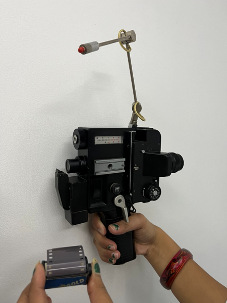
Kowa hand-held fundus camera — the beginning of our imaging journey
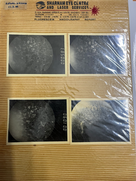
Fluorescein angiography images obtained with the hand-held camera
I still remember that in 1994, my mentor the late Padma Bhushan Dr Badrinath visited my clinic. He was pleased to see how we had improvised the mounting of the hand-held camera and was impressed with the quality of the angiography pictures we were producing with such a simple setup.
Those were different times. We had limited technology, but perhaps more importantly, we had a lot of curiosity and enthusiasm.
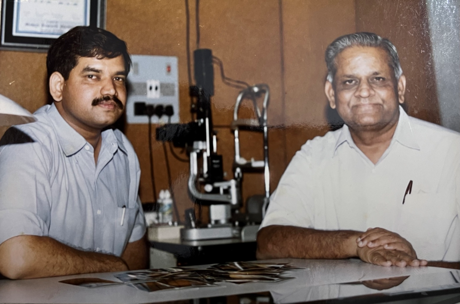
Dr Badrinath viewing the photographs at the clinic, 1994
The Nikon 505 — our next step
In July 1997, we purchased a new Nikon 505 fundus camera for fundus photography and fluorescein angiography.
It was an excellent system and produced very good-quality images. But it was still a film-based camera.
Reporting angiography was therefore a time-consuming process. We actually created a small dark room at our centre where we developed the black-and-white angiography films ourselves. Colour photographs were sent to a nearby colour photo laboratory for printing.
Looking back, it sounds almost primitive compared with today’s instant digital imaging. But at that time, this was how we worked—and we were happy to do whatever was needed to improve diagnosis.
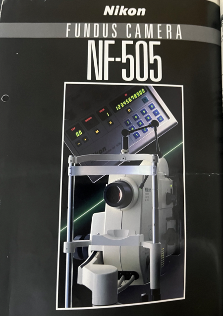
Nikon NF-505 fundus camera brochure
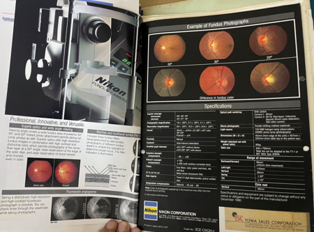
Nikon NF-505 brochure — fundus photography and fluorescein angiography
The digital revolution — Zeiss FF 450 Plus
As technology progressed, film cameras gradually gave way to digital imaging.
In July 2005, we acquired the Zeiss FF 450 Plus fundus camera.
This was a major change for us. The image quality was excellent, and the digital software made image acquisition, storage and interpretation much easier.
For the first time, we could give patients an almost immediate angiography report instead of waiting for films to be developed and photographs printed.
It was not just a new machine. It changed our workflow and changed the way we looked at retinal disease.
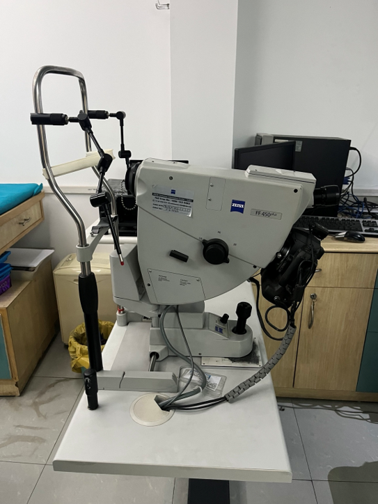
Zeiss FF 450 Plus digital fundus camera
Then came OCT — looking inside the retina
The next major transformation came in June 2006, when we brought a Spectral OCT system from Optopol Germany to Sharnam Eye Centre.
This was perhaps an even bigger change than the transition from film to digital photography.
Until then, much of our understanding of retinal disease came from looking at the surface of the retina through fundus photography and angiography.
OCT allowed us to look at the cross-sectional architecture of the retina.
It was almost like having a biopsy of retinal tissue—without touching the tissue.
We could see the individual retinal layers, fluid, structural changes and subtle abnormalities that could not be appreciated from a photograph alone.
This changed our understanding of retinal diseases and, consequently, our diagnostic and treatment protocols.
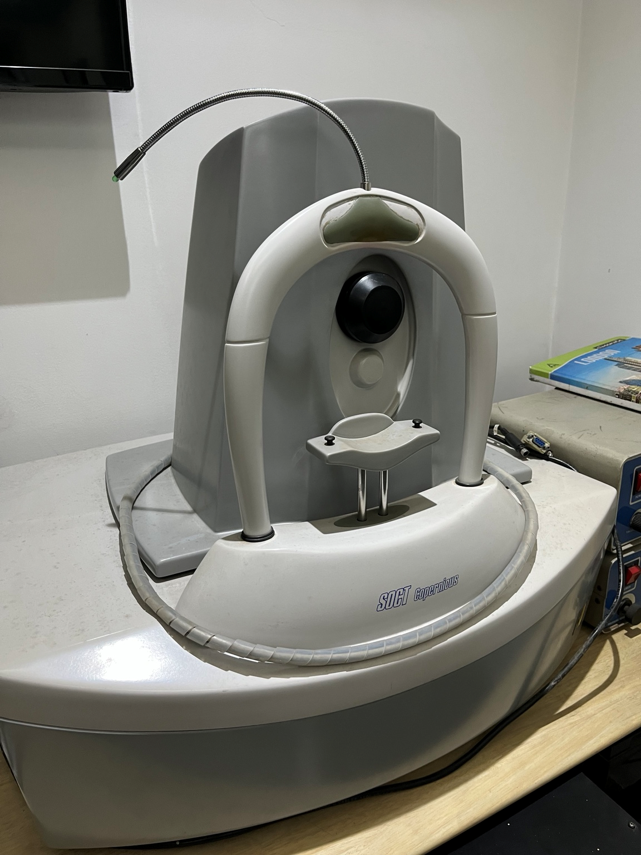
Retinal OCT imaging — the transition to cross-sectional retinal imaging
OCT keeps evolving – OCT Angiography: The Era of Dye-less Angiography
OCT technology did not stop there.
Acquisition became faster and faster. The images became more detailed. Eventually, OCT systems became capable of providing an almost three-dimensional understanding of retinal structure and surface anatomy.
Then came another fascinating development—OCT Angiography.
To be able to see the retinal and choroidal vascular network without injecting dye, and to visualise even very fine capillary networks in the macula, was remarkable.
The macula is one of the finest and most delicate structures in the human body. Being able to study its microvascular architecture non-invasively opened another dimension in retinal diagnosis.
In 2021, we acquired a high-end Heidelberg OCT/OCT Angiography system from Germany.
OCT and OCTA have now become an integral part of our retinal practice.
But interestingly, despite all these advances, fluorescein angiography and conventional retinal photography have not become obsolete. They have continued to evolve alongside OCT and OCTA and remain extremely valuable in many retinal disorders.
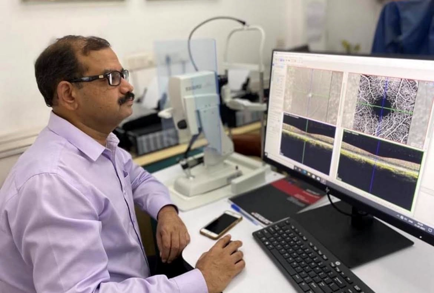
OCT Angiography in clinical practice
Looking further into the periphery
There was another area that continued to fascinate us—the extreme peripheral retina.
The older fundus cameras had a limited field of view. We could examine the periphery clinically with an indirect ophthalmoscope, and this remained extremely useful for identifying peripheral retinal degenerations, tears and other pathology.
In 2025, we added a newer Keeler Video indirect ophthalmoscope system, further strengthening our ability to examine the peripheral retina.
But the desire to photograph what we could see clinically continued.
With the development of wide-field imaging systems, it became possible to capture retinal pathology much farther into the periphery than was possible with conventional fundus cameras.
And this was important because sometimes the most significant pathology is not sitting in the macula—it is hiding somewhere in the periphery.
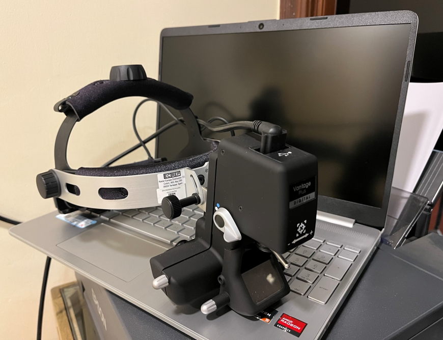
Keeler video indirect ophthalmoscope — imaging the extreme periphery
Zeiss Clarus — bringing the extreme periphery into the picture
Our latest step has been the acquisition of the Zeiss Clarus wide-field imaging and angiography system in July 2026.
This gives us high-quality colour retinal imaging and wide-field angiography, allowing us to document pathology much farther into the retinal periphery.
Wide-field imaging has opened another window into diseases such as diabetic retinopathy, retinal vascular disorders, inflammatory diseases, retinal detachments and peripheral retinal pathology.
Things that were previously described in words or seen only clinically can now be photographed, documented, compared and followed over time.
Taking retinal imaging to the patient
Our imaging journey has not been limited to the equipment inside the centre.
We also wanted to take retinal screening to people who may not be able to come to a specialised retinal centre.
This led us to develop our mobile diabetic retinopathy screening service.
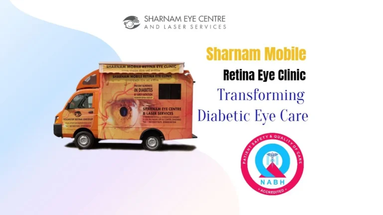
Our mobile retina clinic is equipped with a fundus camera. Images can be captured at the patient’s doorstep or at a physician’s clinic and sent to our centre for evaluation and reporting.
In this way, technology is not merely helping us see better—it is helping us reach more patients.
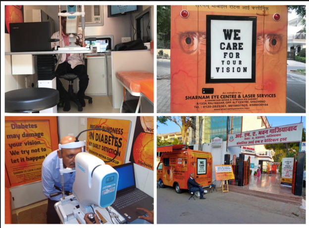
Door step Diabetic Retinopathy screening through Digital Fundus Camera
From 20 degrees to the extreme periphery
When I look at the journey, the transformation is extraordinary.
We started with a 20-degree, film-based, hand-held camera mounted on a table.
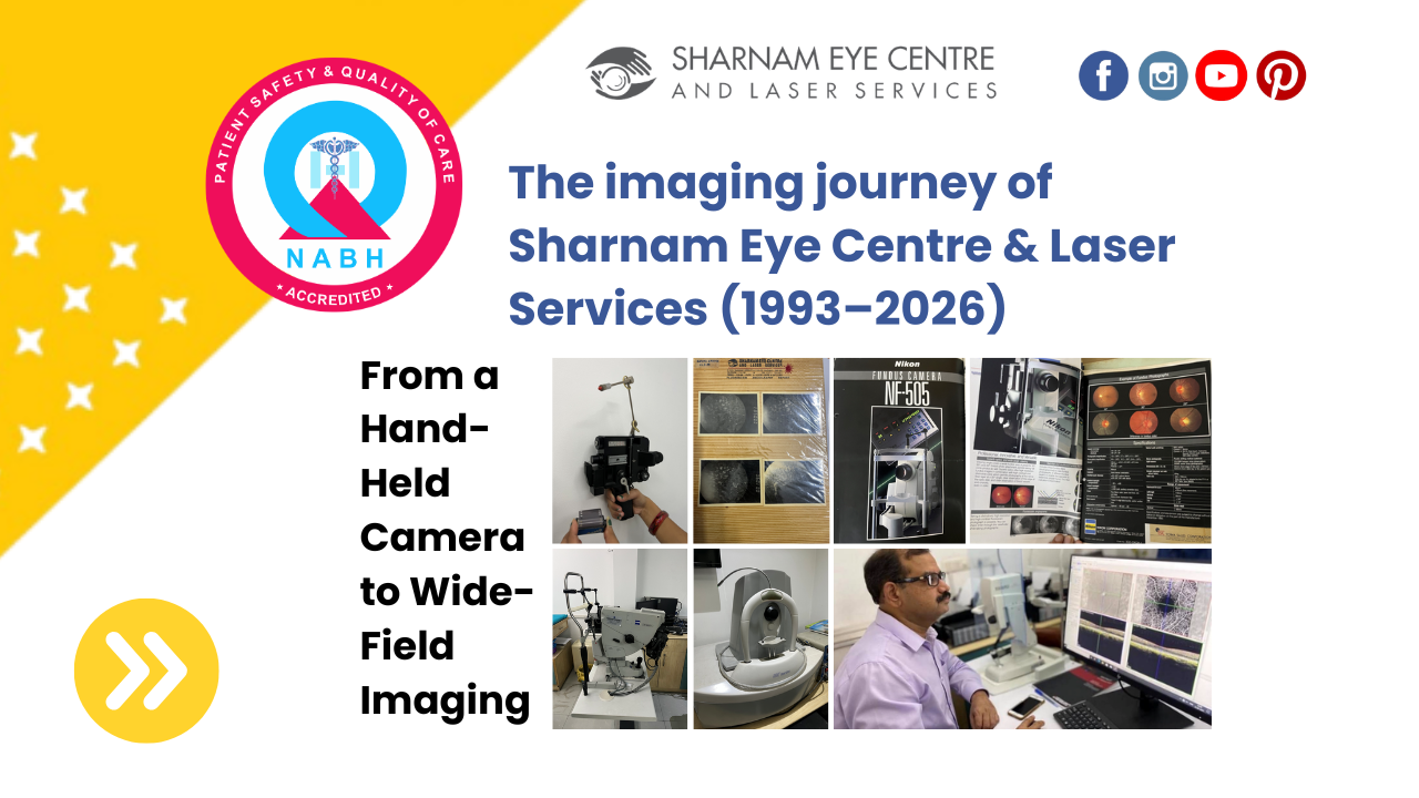
We then moved to Nikon film photography, developed films ourselves, printed colour photographs at a nearby laboratory, moved to digital Zeiss imaging, introduced OCT, progressed to OCT Angiography and finally to wide-field imaging and angiography.
The technology has changed enormously.
But the basic objective has remained exactly the same:
To see better, diagnose better and treat our patients better.
Retina is perhaps one of the most exciting fields in ophthalmology because we can actually see what we treat. Every new imaging technology gives us another way of understanding the retina.
And the journey is far from over.
For me, this story is not really about machines and cameras. It is about curiosity, continuous learning and the willingness to keep moving with technology.
The old Kowa camera, the Nikon 505, the Zeiss FF 450, our first OCT and all the systems that followed are more than pieces of equipment. They are milestones in our journey.
Looking at these old photographs brings back memories of how far retinal imaging has travelled—and how fortunate we are to have witnessed this transformation.
Retina keeps evolving. Imaging keeps evolving. And perhaps that is what keeps us young in the field—never stopping, never assuming that we know enough, and always wanting to see a little more.
Keep learning. Keep adapting. Keep looking. There is always something new to see.