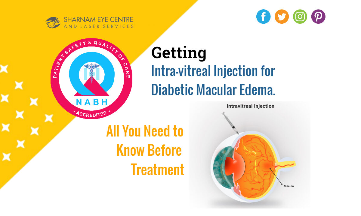
Getting Macular Hole Surgery Done

Getting Macular Hole Surgery Done? All You Need To Know About The Treatment

Macular holes can have a significant impact on your vision, making it crucial to understand the treatment options available. Macular hole surgery is a highly effective procedure that can help restore your vision and improve your quality of life. In this guide, we’ll delve into everything you need to know about macular hole surgery, from the procedure itself to recovery and potential risks.

What Is a Macular Hole?

Before we dive into the surgical aspect, let’s first understand what a macular hole is. The macula is a small, highly sensitive area in the center of the retina responsible for sharp, detailed vision. A macular hole is a small break or tear in this area, leading to distorted or blurred central vision. Macular holes can be caused by a variety of factors, including age-related changes and eye injuries.

Indications for Surgery:

Macular holes are classified into different stages, and not all may require surgery. Your eye doctor will determine the best course of action based on the severity and size of the hole. Typically, surgery is recommended for:

1. Full-thickness macular holes: These are more advanced and tend to require surgical intervention.
2. Holes causing significant vision loss: If the macular hole is impairing your central vision, surgery becomes more critical.

The Surgical Procedure:

Macular hole surgery is formally known as vitrectomy with membrane peeling. It involves several steps:

1. Anesthesia: Local or general anesthesia is administered to ensure you’re comfortable throughout the procedure.
2. Vitrectomy: The surgeon removes the vitreous gel from the center of the eye, allowing access to the macular hole.
3. Membrane Peeling: The delicate membrane overlying the macular hole is gently peeled away, allowing the hole to be better visualized and repaired.
4. Gas or Oil Bubble Placement: Depending on the surgeon’s preference, a gas or oil bubble is injected into the eye to help flatten and close the macular hole. This bubble stays in the eye for a specific duration to facilitate healing.
5. Post-operative Positioning: You may be required to maintain a specific head position for a period to ensure the bubble is in contact with the macula.

Recovery and Rehabilitation:

After macular hole surgery, you’ll need to follow specific guidelines for a successful recovery:

1. Rest: Rest is crucial to allow the eye to heal properly.
2. Eye Drops: Your doctor will prescribe eye drops to prevent infection and reduce inflammation.
3. Head Positioning: Following your surgeon’s instructions for head positioning is essential to ensure the bubble effectively closes the hole.
4. Follow-up Visits: Regular follow-up appointments with your eye specialist are necessary to monitor your progress.

Potential Risks and Complications:

Like any surgical procedure, macular hole surgery carries some risks, including:

1. Infection
2. Retinal detachment
3. Cataracts
4. Elevated eye pressure

Your surgeon will discuss these risks with you and explain how they can be minimized.

Macular hole surgery can be a life-changing procedure for those suffering from this vision-threatening condition. It’s essential to consult with an experienced Retina specialist or Vitreo Retinal surgeon or Trained Ophthalmologist who can assess your specific case and recommend the most suitable treatment plan. With proper care and adherence to post-operative instructions, you can significantly improve your vision and regain a better quality of life.

Disclaimer: This content is for informational purposes only and should not replace professional medical advice, diagnosis, or treatment.

Vitrectomy Surgery for Diabetic Vitreous Haemorrhage

Getting Vitrectomy Surgery for Diabetic Vitreous Haemorrhage ? All You Need to Know Before Getting Treatment

Vitrectomy surgery for diabetic vitreous hemorrhage is a procedure to clear blood and other abnormal tissue from the middle of the eye. If you or someone you know is considering this treatment, here’s what you need to know:

1. What is Vitrectomy?

Vitrectomy is a surgical procedure that involves removing the vitreous gel from the middle of the eye. This is done to clear blood and any scar tissue that may be contributing to vision problems.

2. Why is it Done?

In patients with diabetes, blood vessels can become weak and bleed into the vitreous gel, causing what’s known as a vitreous hemorrhage. This can blur vision. Vitrectomy is performed to restore vision by removing this blood.

3. How is the Procedure Performed?

The surgeon makes small incisions in the white part of the eye. Specialized instruments are then used to remove the vitreous gel and any blood or scar tissue. The eye may be filled with a saline solution, gas, or silicone oil to help it maintain its shape.

4. What are the Risks?

Like all surgeries, vitrectomy has risks. They include:

• Infection
• Bleeding inside the eye
• Retinal detachment
• Increased or decreased eye pressure
• Cataract progression
• Vision loss

5. Recovery:

Post-surgery, patients are usually advised:

• To wear an eye patch for a day or so.
• Avoid strenuous activities and heavy lifting.
• Use prescribed eye drops to prevent infection and reduce inflammation.
• If gas was used, you might be advised to maintain a certain head position for several days.

6. Outcomes:

Many patients experience improved vision after vitrectomy. However, the success largely depends on the overall health of the eye and the severity of the diabetic retinopathy.

7. Alternatives:

Vitrectomy is usually recommended when other treatments, such as laser therapy, don’t work. It’s crucial to discuss all your options with an ophthalmologist.

8. Pre-operative Considerations:

• Control blood sugar levels: It’s vital to have diabetes under control before undergoing the surgery.
• Medication: Inform your doctor about all the medications you take, as some may need to be adjusted before surgery.

9. Post-operative Considerations:

• Vision might be blurry for a few days to weeks after surgery.
• Avoid rubbing your eyes.
• Attend all follow-up appointments to ensure proper healing.

It’s crucial to have a comprehensive discussion with your Retina specialist before deciding on vitrectomy to understand all aspects of the procedure, recovery, and potential risks.

Disclaimer: This content is for informational purposes only and should not replace professional medical advice, diagnosis, or treatment.

Retinal Detachment Surgery

Getting Retinal Detachment Surgery? All You Need to Know Before Getting Treatment

Retinal detachment is a serious eye condition that can lead to vision loss if not treated promptly. If you or a loved one is considering retinal detachment surgery, you probably have numerous questions. In this blog ,we’ll walk you through everything you need to know.

1. Understanding Retinal Detachment:

The retina is a thin layer of tissue at the back of the eye, responsible for receiving light and sending signals to the brain. When the retina detaches from the supportive tissues, it can become deprived of oxygen, leading to potential vision loss.

2. Causes and Symptoms:

Causes: Trauma to the eye, aging, tumors, certain eye diseases like myopia , or inflammation can cause the retina to detach.
Symptoms: Look out for sudden flashes of light, a significant increase in floaters, or a shadow in your peripheral vision. If you experience these, consult an preferably a Retina specialist /or an Ophthalmologist immediately.

3. Types of Retinal Detachment Surgery

There are mainly three types:

Pneumatic Retinopexy: A gas bubble is injected into the eye, which pushes the retina back into place.
Scleral Buckling: A silicone band is placed around the eye, pushing the sclera closer to the detached retina.
Vitrectomy: The vitreous gel is removed from the eye and replaced with a gas bubble or silicone oil.

4. Preparation for Surgery:

Before the procedure:

  • Discuss any medications you’re currently taking with your surgeon.
  • You may need to stop certain medicines a week prior.
  • Ensure you have someone to drive you home post-surgery.

5. Recovery and Post-Op Care

  • You’ll likely wear an eye patch for a day or two.
  • Some surgeries may require you to position your head in a specific way for optimal healing.
  • Regular follow-ups with your surgeon are crucial.

6. Potential Risks:

Every surgery carries some risks. Possible complications include infection, bleeding, high or low eye pressure, or the detachment recurring. However, with skilled surgeons and proper care, these risks are minimized.

7. Success Rates:

While success rates can vary based on the type of detachment and the surgery used, most surgeries have a success rate of 80-90 % .Every patient and eye condition is different , it is essential to have discussion with your Retina specialist regarding the outcome of the surgery .Most patients can expect an improvement in their vision after a succesful reattachnent surgery .

If you suspect you have a retinal detachment or have been recommended for surgery, understanding the process can help alleviate anxieties. Always consult with a Retina specialist / Vitreo Retinal Surgeon / or a trained ophthalmologist to discuss your options and ensure the best possible outcome.

Disclaimer: This content is for informational purposes only and should not replace professional medical advice, diagnosis, or treatment.

What to expect during Retina Examination

What to expect during Retina Examination: Your Step-by-Step Guide

If you’ve been scheduled for a retina examination, it’s natural to have questions about what to expect. A thorough retina examination is crucial for assessing the health of your retinas and ensuring your overall eye wellness. In this blog, we will provide you with a step-by-step guide on what to expect during a retina examination to help you feel more informed and at ease.

1. Comprehensive Eye History:

The first step of your retina examination is typically a discussion of your medical history. Your eye care specialist will ask about any pre-existing conditions, family history of eye diseases, medications you’re taking, and any symptoms you’ve been experiencing.

2. Visual Acuity Test:

You’ll undergo a visual acuity test to assess how well you can see at different distances. This usually involves reading letters or symbols from an eye chart.

3. Pupil Dilation:

To get a clear view of your retinas, your pupils will be dilated using eye drops. This process can take about 15-30 minutes to fully take effect.

4. Intraocular Pressure Measurement:

Your eye pressure may be measured using a device called a tonometer. Elevated eye pressure could be a sign of glaucoma, which can affect the retina.

5. Slit-Lamp Examination:

You will sit in front of a slit lamp, which is a specialized microscope equipped with a bright light. The eye care specialist will examine the front and back of your eyes, including the retina, using this instrument.

6. Retinal Imaging:

High-resolution retinal imaging may be done to capture detailed images of your retinas. This can include Optical Coherence Tomography (OCT) or fundus photography.

7. Visual Field Test:

Some retina examinations include a visual field test to assess your peripheral vision. This is particularly important for conditions like glaucoma.

8. Discussion of Findings:

After the examination, your eye care specialist will discuss their findings with you. They will explain any issues detected, if present, and recommend appropriate treatment or further tests if necessary.

9. Post-Dilation Recovery:

Since your pupils will remain dilated for a few hours, you might be sensitive to light. It’s advisable to bring sunglasses and have someone accompany you for the journey home if needed.

10. Follow-Up Plan:

Depending on the results of your retina examination, your specialist will outline a follow-up plan. This may include treatment, future appointments, or lifestyle adjustments.

Conclusion:

A retina examination is a vital part of maintaining good eye health. Knowing what to expect can help ease any anxiety you may have about the process. Regular retina examinations are essential for early detection and treatment of retinal issues, ensuring your continued enjoyment of clear and healthy vision. If you have any concerns or questions about your retina examination, don’t hesitate to discuss them with your eye care specialist; they are there to provide you with the best possible care and information.

Disclaimer: This content is for informational purposes only and should not replace professional medical advice, diagnosis, or treatment.

The Role of Nutrition in Retina Health

Nourishing Your Vision: The Role of Nutrition in Retina Health

Maintaining good eyesight requires more than just regular check-ups; it also depends on the nutrients you provide your eyes. Your retinas, the light-sensitive tissues at the back of your eyes, play a crucial role in vision, and nutrition plays a vital part in keeping them healthy. In this blog, we will explore the significant role of nutrition in retina health and provide insights into the foods that can benefit your eyes.

Understanding the Retina:

Before we dive into nutrition, let’s briefly understand the importance of the retina. It’s like the camera film of your eye, capturing light and transmitting visual information to your brain. Any damage to the retina can affect your vision, making it essential to maintain its health.

Essential Nutrients for Retina Health:

1. Vitamin A: This vitamin is a key component of rhodopsin, a protein in the retina that helps you see in low light conditions. Foods rich in vitamin A include carrots, sweet potatoes, and spinach.

2. Lutein and Zeaxanthin: These antioxidants help protect the retina from harmful light and oxidative damage. Leafy greens, corn, and egg yolks are good sources.

3. Omega-3 Fatty Acids: Found in fatty fish like salmon, mackerel, and trout, omega-3s help maintain the structural integrity of the retina.

4. Vitamin C: This antioxidant is essential for overall eye health and can be found in citrus fruits, strawberries, and bell peppers.

5. Vitamin E: Nuts, seeds, and spinach are rich in vitamin E, which helps protect the cells in your retina.

6. Zinc: Zinc plays a role in the formation of visual pigments in the retina. It’s found in foods like red meat, poultry, and beans.

Tips for a Retina-Boosting Diet:

  • Incorporate a variety of colorful fruits and vegetables into your meals to ensure a broad spectrum of vitamins and antioxidants.
  • Choose whole grains over refined grains to maintain steady blood sugar levels, which is crucial for retina health.
  • Consume fish rich in omega-3 fatty acids at least twice a week.
  • Limit your intake of processed foods and sugary snacks, as they can negatively impact eye health.

Conclusion:

The saying “you are what you eat” holds true for your eyes as well. By incorporating a balanced and nutrient-rich diet into your lifestyle, you can significantly contribute to the health of your retinas and maintain clear and vibrant vision. However, remember that nutrition is just one aspect of eye care. Regular eye exams with a qualified eye care professional are crucial for detecting and addressing any issues with your retinas early on. With proper care and a nourishing diet, you can look forward to a lifetime of healthy vision.

Disclaimer: This content is for informational purposes only and should not replace professional medical advice, diagnosis, or treatment.

Expert Eye Care Tips

Maintaining Healthy Retinas: Expert Eye Care Tips

Your retinas are essential for clear vision, and taking care of them should be a top priority. Fortunately, there are several simple yet effective ways to maintain healthy retinas and preserve your eyesight. In this blog, we’ll explore expert tips for eye care that can help you ensure the well-being of your retinas.

1. Eat a Nutrient-Rich Diet:

A balanced diet rich in vitamins and minerals is crucial for retina health. Include foods like leafy greens, carrots, fish, and nuts in your diet, as they contain nutrients like lutein and omega-3 fatty acids that promote retinal health.

2. Protect Your Eyes from UV Rays:

Prolonged exposure to UV rays can damage the retinas. Whenever you’re outdoors, wear sunglasses that offer UV protection to shield your eyes from harmful sunlight.

3. Maintain Healthy Blood Sugar Levels:

High blood sugar can lead to conditions like diabetic retinopathy, which can harm your retinas. If you have diabetes, closely monitor your blood sugar and follow your healthcare provider’s recommendations.

4. Manage Blood Pressure and Cholesterol:

Hypertension and high cholesterol levels can affect retinal blood vessels. Regular exercise, a heart-healthy diet, and medication if needed can help manage these risk factors.

5. Quit Smoking:

Smoking increases the risk of macular degeneration and other eye diseases. Quitting smoking can significantly improve your retina’s health and overall eye wellness.

6. Exercise Regularly:

Regular physical activity improves blood circulation, which is essential for healthy retinas. Aim for at least 150 minutes of moderate-intensity exercise per week.

7. Maintain a Healthy Weight:

Being overweight or obese can increase the risk of diabetes and other health conditions that affect the retina. Maintain a healthy weight through diet and exercise.

8. Stay Hydrated:

Proper hydration ensures that your eyes receive the necessary nutrients and oxygen. Drink an adequate amount of water daily to support eye health.

9. Protect Your Eyes During Work and Hobbies:

If your job involves prolonged computer use, take regular breaks to reduce eye strain. When engaging in hobbies like reading or crafting, ensure proper lighting to prevent eye fatigue.

10. Schedule Regular Eye Exams:

Routine eye exams with a qualified eye care professional are essential for detecting retinal issues early. Early diagnosis and treatment can prevent the progression of eye diseases.

Conclusion:

Your retinas are irreplaceable, and taking proactive steps to care for them is essential. By adopting these expert-recommended eye care tips, you can significantly reduce the risk of retinal problems and maintain clear and healthy vision for years to come. Remember, a commitment to eye health today ensures a brighter and clearer tomorrow.

Disclaimer: This content is for informational purposes only and should not replace professional medical advice, diagnosis, or treatment.

रेटिना स्पेशलिस्ट या रेटिना विशेषज्ञ क्या होता है?

रेटिना स्पेशलिस्ट या रेटिना विशेषज्ञ क्या होता है?

जिस ऑफ्थाल्मोलॉजिस्ट या नेत्र विशेषज्ञ ने रेटिना की बीमारियों के इलाज में विशेष ट्रेनिंग हासिल की होती है। वह आँख के अंदरूनी भाग रेटिना के बीमारियों और उपचार में माहिर होता है। रेटिना स्पेशलिस्ट जो रेटिना की सर्जरी करते हैं उन्हें विट्रियो रेटिनल सर्जन भी कहा जाता है । यह अपने आप में एक विशेष ब्रांच है जिसमें रेटिना से संबंधित जटिल रोगों के इलाज की दक्षता प्राप्त होती है।

रेटिना क्या है?

रेटिना आँख के पीछे का वह हिस्सा है जहाँ प्रकाश की किरणें पड़कर हमें दृश्य दिखाई देती हैं। यह जब कार्यक्षम नहीं होता, तो दृश्य समस्याएं उत्पन्न होती हैं।

रेटिना स्पेशलिस्ट क्यों जरूरी हैं?

रेटिना से संबंधित समस्याएं जैसे डायबिटिक रेटिनोपैथी, मैकुलर डिजेनेरेशन , रेटिना डिटेचमेंट , मैकुलर होल, वैस्कुलर आक्लूज़न , सेंट्रल सिरस रिटिनोपैथी, रेटिनाइटिस पिग्मेंटोसा आदि जटिल बीमारियों हो सकती हैं और यदि इसे समय पर नहीं पहचाना जाए तो यह दृश्य की हानि का कारण बन सकता है।

कैसे पहचानें कि आपको रेटिना स्पेशलिस्ट की जरूरत है?

यदि आपको धुंधला दिखाई दे रहा है, या आपके दृश्य में अचानक बदलाव महसूस हो रहा है, तो आपको तुरंत रेटिना स्पेशलिस्ट से संपर्क करना चाहिए।आमतोर से नेत्र विशेषज्ञ रेटिना की बीमारी के होने पर रेटिना विशेषज्ञ को रेफ़र भी करते हैं ।

रेटिना स्पेशलिस्ट विशेषज्ञ डॉक्टर हैं जो आँख के रेटिना से संबंधित समस्याओं का समाधान प्रदान करते हैं। अगर आपको लगता है कि आपकी आँखों में कोई समस्या है, तो तुरंत चिकित्सक की सलाह लें।

Disclaimer: This content is for informational purposes only and should not replace professional medical advice, diagnosis, or treatment.

Role of Regular Eye Exams in Preventing Retina Problems

The Crucial Role of Regular Eye Exams in Preventing Retina Problems

Your eyes are your windows to the world, and preserving your vision is of utmost importance. One of the most effective ways to safeguard your eye health, especially concerning retina-related issues, is through regular eye exams. In this blog, we’ll delve into the significance of these exams in preventing retina problems and maintaining your overall visual well-being.

Why Are Regular Eye Exams Necessary?

1. Early Detection of Retina Problems: Regular eye exams enable early detection of retina problems, even before noticeable symptoms appear. Conditions like diabetic retinopathy, macular degeneration, and retinal detachments can be diagnosed and treated promptly, preventing further complications.

2. Monitoring High-Risk Groups: If you have a family history of eye diseases, diabetes, or are over the age of 40, you are at a higher risk of developing retina issues. Regular eye exams ensure close monitoring of high-risk individuals, increasing the chances of timely intervention.

3. Preserving Your Sight: The retina plays a vital role in vision, capturing light and transmitting visual information to the brain. Detecting and addressing retina problems early can significantly impact the preservation of your sight, allowing you to enjoy a better quality of life.

What Happens During an Eye Exam?

During a comprehensive eye exam, your eye care specialist or optometrist will:

  • Assess your visual acuity (clarity of vision).
  • Examine the retina using specialized instruments to detect abnormalities.
  • Check your eye pressure to screen for glaucoma.
  • Evaluate the health of the blood vessels in the retina, crucial for conditions like diabetic retinopathy.

When Should You Get Regular Eye Exams?

The frequency of eye exams can vary based on your age and risk factors:

  • Adults under 40 with no risk factors: Every 2-3 years.
  • Adults over 40: Annually or as recommended by your eye care professional.
  • High-risk individuals: As advised by your eye doctor.

Conclusion:

In conclusion, regular eye exams are not just routine check-ups; they are your shield against potential retina problems that could compromise your vision. Early detection through these exams is the key to preventing and managing retina-related conditions effectively. Your eyesight is invaluable, and by scheduling regular eye exams, you take a proactive step in preserving it for a lifetime of clear and vibrant vision. Don’t wait until a problem arises; prioritize your eye health today for a brighter tomorrow.

Disclaimer: This content is for informational purposes only and should not replace professional medical advice, diagnosis, or treatment.

Understanding Common Retina Diseases: Causes, Symptoms, and Treatments

Your vision is a precious gift, and the retina plays a crucial role in maintaining it. The retina is a thin layer of tissue at the back of your eye that captures light and sends visual signals to your brain. However, like any part of the body, the retina can be affected by various diseases. In this blog, we will explore some of the common retina diseases, their causes, symptoms, and available treatments.

1. Age-Related Macular Degeneration (AMD):

Causes: AMD is primarily linked to aging and genetic factors.
Symptoms: Blurred or distorted central vision, difficulty reading, and recognizing faces.
Treatments: Depending on the type of AMD, treatments can include medications, laser therapy, or injections.

2. Diabetic Retinopathy:

Causes: High blood sugar levels damage blood vessels in the retina.
Symptoms: Initially, there may be no symptoms, but as the disease progresses, you might experience vision changes.
Treatments: Managing diabetes is essential. Treatments may include laser therapy or injections.

3. Retinal Detachment:

Causes: This occurs when the retina pulls away from its normal position.
Symptoms: Sudden flashes of light, a curtain-like shadow over your vision, or a sudden increase in floaters.
Treatments: Surgery is typically required to reattach the retina.

4. Retinitis Pigmentosa:

Causes: It’s often a genetic disorder.
Symptoms: Night blindness, tunnel vision, and a gradual loss of peripheral vision.
Treatments: While there is no cure, low vision aids and genetic counseling may help manage the condition.

5. Macular Hole:

Causes: Usually occurs due to age or trauma.
Symptoms: Blurred or distorted central vision.
Treatments: Surgical intervention is often necessary to repair the hole.

6. Retinoblastoma:

Causes: A rare cancer that typically affects young children.
Symptoms: White pupil, misaligned eyes, and vision problems.
Treatments: Treatment options include chemotherapy, radiation therapy, or surgery.

Conclusion:

Your retina is a vital component of your vision, and understanding common retina diseases is essential for early detection and treatment. Regular eye exams are key to identifying any issues early on. If you experience any unusual vision changes or symptoms, don’t hesitate to reach out to a retina specialist like Dr. Puneet Gupta, who can provide expert guidance and care tailored to your needs. Remember, early diagnosis and prompt treatment can often make a significant difference in preserving your vision and quality of life.

Disclaimer: This content is for informational purposes only and should not replace professional medical advice, diagnosis, or treatment.

Appointment
close slider

For Appointment-

Online Appointment: Available between 8 AM to 8 PM (Except Saturday)

Book An Online Appointment - Click Here

Call for Appointment -

9818851925,

9810979457,

8586030104

-----------------------------------------