A Heartfelt Gesture: From Macular Degeneration to a Canvas of Gratitude

When you’re in the medical profession, your ultimate reward is often the improved health and happiness of your patients. But every once in a while, a gesture comes along that strikes a profound chord and makes every moment of hard work truly worth it. This is one such story.

Journey from Darkness to Light:

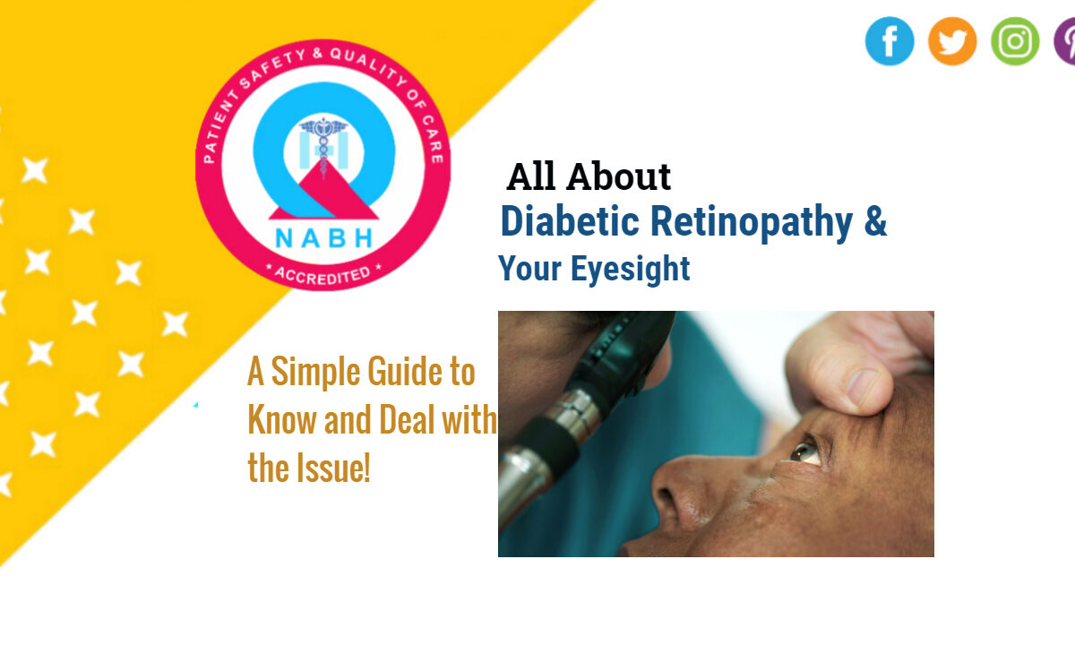
One of my most memorable patients came to our centre with macular degeneration in both eyes. Macular degeneration, for those unfamiliar, is a serious eye condition that can lead to severe vision loss. Faced with the daunting prospect of losing her sight, we decided to administer multiple intravitreal anti-VEGF injections.

The Miracle of Modern Medicine:

Intravitreal anti-VEGF injections have revolutionized the treatment for serious vision-threatening conditions like diabetic retinal disease and, of course, macular degeneration. They target abnormal blood vessel growth in the eyes, preserving and often restoring vision.

To our immense joy, her treatment was successful! She not only regained her vision but experienced a new lease of life.

A Token of Appreciation:

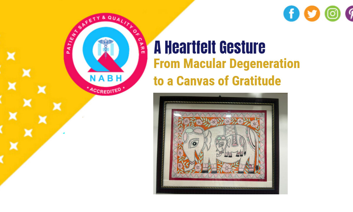
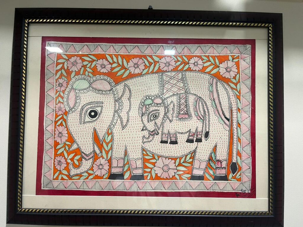
Soon after her recovery, she presented us with a beautiful surprise: a hand-painted piece of art she’d crafted herself. This wasn’t just any painting. It was a testament to her regained vision, a canvas of her journey from darkness to clarity, and a heartfelt token of gratitude.

The Ripple Effect of Gratitude:

Her gesture resonated deeply with me and my entire team. While it might seem like a simple act to some, for us, it signified the profound impact our efforts had on a patient’s life. It’s these moments that rejuvenate our spirits, remind us of our purpose, and inspire us to constantly elevate our standard of care.

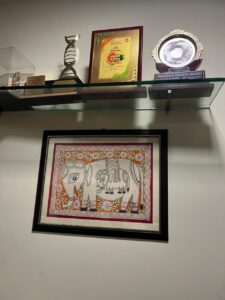
In the vast world of medicine, amidst the technicalities and the challenges, it’s essential to remember the human element – the bonds formed, the stories shared, and the lives changed. This painting, hanging proudly in our centre, serves as a daily reminder of the impact we can have and the gratitude that sometimes comes full circle.


While we always strive for excellence in our services, it’s these unsolicited gestures of appreciation that drive home the point that healthcare is as much about human connection as it is about healing. And as we continue to provide care, it’s these moments of heartfelt gratitude that motivate us to keep pushing boundaries and making a difference.

All About Diabetic Retinopathy and Your Eyesight

Diabetic Retinopathy and Your Eyesight – A Simple Guide to Know and Deal with the Issue!

If you find out you have diabetic retinopathy, it can be scary. You might be worried about losing your sight or how to treat it. The good news is that knowing about it and seeing your doctor can help avoid big problems.

What is Diabetic Retinopathy?

Diabetic retinopathy happens when high sugar levels hurt the back part of your eye, called the retina. Some people don’t know they have it because one eye might help the other see better.

Why Eye Check-ups Matter:

Seeing your Retina eye doctor often is super important. Dr. Puneet Gupta, an Retina expert, says that if you catch it early, you can stop a lot of the damage. If you’ve had diabetes for a long time, you should see your Retina specialist /eye doctor more often. And if you notice things like floaty spots, blurry vision, or colors looking faded, call your eye doctor right away.

Can You Get Better If You Lose Sight?

If you take good care of your health—like watching your sugar levels, eating right, not smoking, and taking your medicine—you might get some of your sight back. This is not only good for your eyes but also for your whole body.

How Does It Hurt Your Eyes?

The damage can be different for everyone. At first, tiny blood vessels in your eye might leak or get puffy. Later on, weak new blood vessels can grow and bleed inside the eye. In the worst cases, these blood vessels make scars that can pull the retina away from the eye.

Treatments Today:

There are good treatments nowadays for diabetic retinopathy. Some people might get medicine shots / injections in the eye or laser treatments. Some might need a surgery called vitrectomy. If the idea of a injection in your eye scares you, don’t worry. It’s usually fast and doesn’t hurt much.

Life with Less Sight:

If you do lose some sight, you can still have a good life. There are tools like magnifying glasses, talking devices, and apps to help .

To sum it up, having diabetic retinopathy is serious, but with the right care and early action, you can handle it. Always remember to see your Retina specialist or eye doctor, ask questions, and get the help you need.

Dr. Puneet Gupta’s Enlightening Talk on OCT Angiography in Bareilly

On October 14th, 2023, the Bareilly Ophthalmological Society held a event that saw a gathering of many of the city’s renowned ophthalmologists. The highlight of this event was a talk presented by Dr. Puneet Gupta, the Director of Sharnam Eye Centre and Laser Services.

The main focus of Dr. Gupta’s presentation was on OCT Optical Coherence Tomography Angiography and its clinical significance in age-related macular degeneration. OCT angiography, a non-invasive imaging Dye less angiography technique of retina , offers insights into the retinal layers and blood vessels, playing an essential role in modern ophthalmology.

Dr. Gupta’s interest in OCT and OCT angiography, along with his expertise in minimal invasive vitreo retinal surgery, made him a fitting speaker for the topic. The attendees found the session to be informative, with many ophthalmologists from Bareilly actively participating and discussing their experiences and queries related to the subject.

The interactive nature of the session facilitated a fruitful exchange of knowledge and perspectives. The Bareilly Ophthalmological Society’s decision to invite Dr. Gupta for the talk reflected the relevance and timeliness of the topic in the ever-evolving field of ophthalmology.

To sum it up, the event served as a platform for professionals to gain deeper insights into OCT angiography and its implications. With experts like Dr. Puneet Gupta sharing their knowledge, the medical community continues to stay updated on the latest advancements in eye care.

World Sight Day Celebrations At Sharnam Eye Centre

World Sight Day Celebrations at Sharnam Eye Centre and Laser Services, Ghaziabad

On 12th October, the world observed ‘World Sight Day’, a day dedicated to the cause of preventing blindness and promoting vision care. Our eye center, Sharnam Eye Centre and Laser Services, was no exception to these celebrations.

The day started bright and early at Central Park, Rajnagar, a favored haunt for morning walkers in the city. Here, Sharnam’s Mobile Retina Clinic came alive with activity as it began retinal imaging of diabetic patients. Diabetic retinopathy screening – a crucial procedure for those with diabetes – was conducted smoothly, drawing many to benefit from this vital service. Many participants, enthusiastic about the cause, posted selfies with the World Sight Day banner at the mobile van, spreading awareness and showing their support for the cause.

In addition to the screenings, attendees were provided with a digital eye care tips brochure, equipping them with knowledge on how to better care for their vision. This digital resource proved popular among the crowd, ensuring that the event’s impact lasted beyond just one day.

Back at the Sharnam Eye Centre, patients too were not left out of the celebrations. Many took the opportunity to pose with the World Sight Day standee placed strategically within the premises, after which they too received the eye care tips digital brochure.

This year’s theme, ‘Love Your Eyes at Work,’ addressed a pressing issue of our times – digital eye strain. With the predominance of screen-based tasks in today’s work culture, many face the brunt of this ailment. The brochure provided helpful tips tailored to help those suffering from or at risk of digital eye strain.

As evening approached, the Sharnam mobile retina clinic found its place in front of the popular Gaur Mall in the RDC area. This move was made possible thanks to a collaboration with the RDC Welfare Association, an organization representing the traders of the RDC area. The evening saw a different demographic, as many young individuals approached the van. Eager to learn, they benefitted immensely from the eye care information provided.

The entire day’s activities were a testament to the dedication of Sharnam Eye Centre and Laser Services towards the vision care cause. It was evident that the team left no stone unturned in ensuring that World Sight Day in Ghaziabad was both informative and engaging. As the day wrapped up, it was clear that the event had achieved its goal, leaving a lasting impact on all those who participated.

2023 World Sight Day

Celebrate World Sight Day 2023 With Sharnam Eye Centre: Love Your Eyes

On October 12, 2023, World Sight Day is celebrated with enthusiasm and commitment at Sharnam Eye Centre and Laser Services, Ghaziabad. This notable centre, specializing in retina and vitreous eye diseases, joins the global initiative to shed light on blindness and vision impairment, emphasizing this year’s theme: “Love Your Eyes at Work”.

Sharnam’s Dedication to Eye Health:

Sharnam Eye Centre stands as a beacon for those seeking quality care and treatment for various eye conditions. With a focus on retina and vitreous diseases, the centre is equipped with state-of-the-art technology and a team of dedicated professionals ready to serve the community in and around Ghaziabad.

World Sight Day 2023 Theme:

World Sight Day 2023 prompts us to pay special attention to our eyes, especially within our work environments. With the theme “Love Your Eyes at Work”, the day aims to raise awareness about the importance of eye health and the preventative measures individuals can take to avoid eye strain and potential vision loss.

World Sight Day 2023

How to Love Your Eyes: 10 Essential Tips

  • Practice the 20/20/20 Rule: Give your eyes the break they need from screens by following the 20/20/20 rule. Every 20 minutes, glance at something 20 feet away for at least 20 seconds. This simple practice helps prevent eye strain and headaches.
  • Embrace the Outdoors: Encourage children to spend a minimum of two hours outdoors daily. Exposure to natural light aids healthy eye development and reduces the risk of nearsightedness.
  • Sport Sunglasses: Whenever you step outside, protect your eyes with sunglasses that offer UVA and UVB protection. Shielding your eyes from harmful solar radiation is crucial for maintaining eye health.
  • Wear Prescription Glasses as Needed: If prescribed, consistently wear your glasses to alleviate strain on your eyes, preventing further complications and ensuring clear vision.
  • Monitor Cosmetics: Be vigilant about the expiration dates on your eye makeup and regularly replace brushes and applicators to prevent bacterial buildup and subsequent infections.
  • Stay Active: Regular exercise not only keeps you fit but also reduces the risk of conditions like diabetes and high blood pressure, which can adversely affect your eyesight.
  • Maintain a Balanced Diet: Fuel your body with a diet rich in essential vitamins and minerals to support and sustain healthy eyes.
  • Say No to Smoking: Avoid smoking to minimize the risk of severe eye conditions and irreversible vision loss.
  • Undergo Regular Eye Examinations: Early detection is key in managing eye conditions effectively. Schedule regular eye examinations to monitor your eye health, even if you haven’t noticed any issues.
  • Prioritize Your Eye Health: Your eyes are meant to last a lifetime, so give them the care and attention they deserve. Regular checks and mindful practices can ensure a lifetime of good vision.

Conclusion:

Sharnam Eye Centre and Laser Services is committed to providing stellar eye care services in Ghaziabad. On this World Sight Day, we urge everyone to not only love your eyes at work but to integrate these practices into your daily routine for lifelong healthy vision. Celebrate this day with us, and let’s work together towards a world where everyone has access to the gift of sight.

Intra-vitreal Injection for Macular Degeneration

Getting Intra-vitreal Injection for Macular Degeneration? All You Need to Know Before Getting Treatment

Macular degeneration is not an uncommon eye disorder, primarily affecting the elderly. If you’ve been recommended an intra-vitreal injection as treatment, it’s essential to be informed. In this guide blog, we’ll walk you through everything you need to know about the procedure.

1. Understanding Macular Degeneration:

Macular degeneration, often referred to as Age-Related Macular Degeneration (AMD), affects the central portion of the retina called the macula. This disorder can lead to blurred or no vision in the center of the visual field. Early diagnosis and treatment are crucial for maintaining optimal vision.

2. What is an Intra-vitreal Injection?

An intra-vitreal injection involves injecting medicine directly into the vitreous, the jelly-like substance in the eye. This procedure delivers the medication right where it’s needed, targeting the affected retina areas.

3. Why Opt for Intra-vitreal Injections?

For certain forms of AMD, notably the wet form, intra-vitreal injections of anti-VEGF medications can slow the disease’s progression. These injections can:

  • Reduce swelling
  • Limit the growth of abnormal blood vessels
  • Improve or stabilize vision

4. Preparing for the Procedure:

Before the injection:

  • Your eye will be cleaned and numbed
  • You might undergo an Optical coherence tomography (OCT) scan or Angiography to determine the retina’s status

5. Aftercare and Recovery:

Post-injection, you may experience:

  • Mild discomfort or a gritty sensation
  • Temporary eye redness

It’s vital to follow your doctor’s aftercare instructions and watch for signs of infection or complications.

6. Potential Side Effects:

Though intra-vitreal injections are generally safe, some potential side effects include:

  • Eye pain or redness
  • Floaters
  • Increased eye pressure
  • Infection

7. How Often Are Injections Needed?

The frequency of injections depends on the medication used and your eye’s response. There are several injections available like Razumab, Accentrix, Pegenex, Eylea , your Retina specialist will choose the best suited for your retina condition . These injections are given based on a protocol which can be tailored as per patients need and condition . You must discuss all the aspects before the treatment is initiated . Some patients might need monthly injections, while others might require less frequent treatments. Since some patients may require more injections to stabilize the disease process one must be patient and keep discussing his condition and treatment outcomes on follow up visits .

8. Cost and Insurance:

Sone newer Intra-vitreal injections can be expensive. Ensure you check with your insurance provider about coverage and co-payments. Financial assistance programs might be available to help with costs . Your retina specialist will help you in choosing the right treatment for you and also help in tailoring the treatment based on your condition and need .

Being informed is your best defense against macular degeneration. An intra-vitreal injection can be an effective treatment, but it’s crucial to understand the procedure, its benefits, and potential risks. Always consult with a Retina specialist or an trained ophthalmologist to discuss your options and get the best care for your eyes.

Disclaimer: This content is for informational purposes only and should not replace professional medical advice, diagnosis, or treatment.

Intra-vitreal Injection for Diabetic Macular Edema

Getting Intra-vitreal Injection for Diabetic Macular Edema: All You Need to Know Before Treatment

Diabetic macular edema (DME) is a common complication in those suffering from diabetes. It involves swelling in the macula due to fluid leakage, which can significantly impact vision. One effective treatment for DME is intra-vitreal injection. If you or someone you know is considering this treatment, here’s a comprehensive guide to ensure you’re well-informed.

1. What is an Intra-vitreal Injection?

Intra-vitreal injection involves injecting medication directly into the vitreous, the jelly-like substance inside your eye. This method ensures direct delivery of the medication to the retina, providing relief from DME symptoms.

2. Why Choose Intra-vitreal Injection for DME?

Several reasons make intra-vitreal injections a preferred choice:

  • Targeted Treatment: Directly addresses the affected area.
  • Fewer Side Effects: Intra vitreal injections are usually safe.
  • Quick Procedure: Takes only a few minutes under local anesthesia.

3. Pre-procedure Preparations:

Before getting the injection, you’ll need a thorough eye examination including OCT or / Angiography . It’s also essential to inform your retina specialist or Ophthalmologist about any medications you’re taking, as some may interact with the DME treatment.

4. What to Expect During the Procedure?

The entire procedure is quick and relatively painless:

  • The eye is numbed using drops.
  • A speculum is used to keep the eye open.
  • The injection is administered with a tiny needle.
  • Antibiotic drops are often prescribed post-procedure.

These injections are given with a very fine needle, under microscope very precisely after numbing the eye hence they are safe and painless .

5. Post-procedure Care:

It’s vital to monitor any changes in your vision after the injection. Regular eye and retina check up along with OCT scan is essential to monitor the recovery and modify the treatment if required . Any sudden decrease in vision or increased redness and pain should be reported immediately.

6. Benefits and Risks:

Like all treatments, intra-vitreal injections come with both benefits and risks. While they can significantly improve vision and reduce swelling, potential risks include infection, retinal detachment, and increased eye pressure.

7. Cost:

The cost can vary depending on the medication used . There are choices like Razumab, Accentrix, Eylea, Pegenex and Ozurdex, your retina specialist will inform and discuss about what is best for you . The treatment is done following a protocol tailored as per patient s need .

Conclusion:

Intra-vitreal injections are an effective solution for DME, providing relief and improving vision. Always consult with an Retina specialist or a trained ophthalmologist to determine if it’s the right treatment for you.

Disclaimer: This content is for informational purposes only and should not replace professional medical advice, diagnosis, or treatment.

Getting Macular Hole Surgery Done

Getting Macular Hole Surgery Done? All You Need To Know About The Treatment

Macular holes can have a significant impact on your vision, making it crucial to understand the treatment options available. Macular hole surgery is a highly effective procedure that can help restore your vision and improve your quality of life. In this guide, we’ll delve into everything you need to know about macular hole surgery, from the procedure itself to recovery and potential risks.

What Is a Macular Hole?

Before we dive into the surgical aspect, let’s first understand what a macular hole is. The macula is a small, highly sensitive area in the center of the retina responsible for sharp, detailed vision. A macular hole is a small break or tear in this area, leading to distorted or blurred central vision. Macular holes can be caused by a variety of factors, including age-related changes and eye injuries.

Indications for Surgery:

Macular holes are classified into different stages, and not all may require surgery. Your eye doctor will determine the best course of action based on the severity and size of the hole. Typically, surgery is recommended for:

1. Full-thickness macular holes: These are more advanced and tend to require surgical intervention.
2. Holes causing significant vision loss: If the macular hole is impairing your central vision, surgery becomes more critical.

The Surgical Procedure:

Macular hole surgery is formally known as vitrectomy with membrane peeling. It involves several steps:

1. Anesthesia: Local or general anesthesia is administered to ensure you’re comfortable throughout the procedure.
2. Vitrectomy: The surgeon removes the vitreous gel from the center of the eye, allowing access to the macular hole.
3. Membrane Peeling: The delicate membrane overlying the macular hole is gently peeled away, allowing the hole to be better visualized and repaired.
4. Gas or Oil Bubble Placement: Depending on the surgeon’s preference, a gas or oil bubble is injected into the eye to help flatten and close the macular hole. This bubble stays in the eye for a specific duration to facilitate healing.
5. Post-operative Positioning: You may be required to maintain a specific head position for a period to ensure the bubble is in contact with the macula.

Recovery and Rehabilitation:

After macular hole surgery, you’ll need to follow specific guidelines for a successful recovery:

1. Rest: Rest is crucial to allow the eye to heal properly.
2. Eye Drops: Your doctor will prescribe eye drops to prevent infection and reduce inflammation.
3. Head Positioning: Following your surgeon’s instructions for head positioning is essential to ensure the bubble effectively closes the hole.
4. Follow-up Visits: Regular follow-up appointments with your eye specialist are necessary to monitor your progress.

Potential Risks and Complications:

Like any surgical procedure, macular hole surgery carries some risks, including:

1. Infection
2. Retinal detachment
3. Cataracts
4. Elevated eye pressure

Your surgeon will discuss these risks with you and explain how they can be minimized.

Macular hole surgery can be a life-changing procedure for those suffering from this vision-threatening condition. It’s essential to consult with an experienced Retina specialist or Vitreo Retinal surgeon or Trained Ophthalmologist who can assess your specific case and recommend the most suitable treatment plan. With proper care and adherence to post-operative instructions, you can significantly improve your vision and regain a better quality of life.

Disclaimer: This content is for informational purposes only and should not replace professional medical advice, diagnosis, or treatment.

Vitrectomy Surgery for Diabetic Vitreous Haemorrhage

Getting Vitrectomy Surgery for Diabetic Vitreous Haemorrhage ? All You Need to Know Before Getting Treatment

Vitrectomy surgery for diabetic vitreous hemorrhage is a procedure to clear blood and other abnormal tissue from the middle of the eye. If you or someone you know is considering this treatment, here’s what you need to know:

1. What is Vitrectomy?

Vitrectomy is a surgical procedure that involves removing the vitreous gel from the middle of the eye. This is done to clear blood and any scar tissue that may be contributing to vision problems.

2. Why is it Done?

In patients with diabetes, blood vessels can become weak and bleed into the vitreous gel, causing what’s known as a vitreous hemorrhage. This can blur vision. Vitrectomy is performed to restore vision by removing this blood.

3. How is the Procedure Performed?

The surgeon makes small incisions in the white part of the eye. Specialized instruments are then used to remove the vitreous gel and any blood or scar tissue. The eye may be filled with a saline solution, gas, or silicone oil to help it maintain its shape.

4. What are the Risks?

Like all surgeries, vitrectomy has risks. They include:

• Infection
• Bleeding inside the eye
• Retinal detachment
• Increased or decreased eye pressure
• Cataract progression
• Vision loss

5. Recovery:

Post-surgery, patients are usually advised:

• To wear an eye patch for a day or so.
• Avoid strenuous activities and heavy lifting.
• Use prescribed eye drops to prevent infection and reduce inflammation.
• If gas was used, you might be advised to maintain a certain head position for several days.

6. Outcomes:

Many patients experience improved vision after vitrectomy. However, the success largely depends on the overall health of the eye and the severity of the diabetic retinopathy.

7. Alternatives:

Vitrectomy is usually recommended when other treatments, such as laser therapy, don’t work. It’s crucial to discuss all your options with an ophthalmologist.

8. Pre-operative Considerations:

• Control blood sugar levels: It’s vital to have diabetes under control before undergoing the surgery.
• Medication: Inform your doctor about all the medications you take, as some may need to be adjusted before surgery.

9. Post-operative Considerations:

• Vision might be blurry for a few days to weeks after surgery.
• Avoid rubbing your eyes.
• Attend all follow-up appointments to ensure proper healing.

It’s crucial to have a comprehensive discussion with your Retina specialist before deciding on vitrectomy to understand all aspects of the procedure, recovery, and potential risks.

Disclaimer: This content is for informational purposes only and should not replace professional medical advice, diagnosis, or treatment.

Appointment
close slider

For Appointment-

Online Appointment: Available between 8 AM to 8 PM (Except Saturday)

Book An Online Appointment - Click Here

Call for Appointment -

9818851925,

9810979457,

8586030104

-----------------------------------------