Diabetic Eye Disease
Diabetic Retinopathy
Nearly half of people with diabetes have some degree of diabetic retinopathy, a complication of diabetes that can result in blindness. After 20 years with diabetes, most patients have diabetic retinopathy. Treatment depends on the severity of the condition
What is Diabetic Retinopathy?
When people have diabetes, excess glucose in the bloodstream can cause the tiny capillaries in the back of the eye to swell and leak fluid. This causes blurry vision. Tiny new blood vessels grow out of the retina as the disease advances. These blood vessels may break and bleed into the vitreous, further impairing vision. Initially, you may not notice changes in vision even though you may have the early stages of diabetic retinopathy. However, as the retinopathy progresses, you may start to notice a decline in vision, usually in both eyes. With advanced retinopathy, you may have severe vision loss or blindness.
What are the stages of diabetic retinopathy?
There are two types of diabetic retinopathy; proliferative diabetic retinopathy is the more serious and should be treated promptly.
Nonproliferative diabetic retinopathy (NPDR): This is the early stage of the disease. Symptoms often are mild or nonexistent. However, blurred vision may occur from swelling of the retina. This swelling occurs because the damaged blood vessels can ooze fluid.
You may not need treatment immediately. Your eye doctor will closely monitor changes in the retina. You need to work with your general medical doctors to control your glucose, blood pressure and cholesterol.
Proliferative diabetic retinopathy (PDR): This is the more advanced form of the disease, where abnormal new blood vessels grow on the surface of the retina or into the vitreous cavity. These new vessels may bleed into the vitreous, clouding or blocking vision. The blood vessels can pull on the retina, leading to retinal detachment.
Macular edema/clinically significant macular edema (CSME): This is responsible for diminition of vision and happens due to leakage of fluid from the damaged blood vessels and needs immediate treatment. It may coexist with either NPDR or PDR.
Who is at risk for diabetic retinopathy and what preventative measures can be taken?
Individuals with Type I, Type II, and gestational (during pregnancy) diabetes are all at risk. As there are no symptoms in the early stages of diabetic retinopathy, individuals with diabetes should have a comprehensive dilated exam once a year.
However, depending on the stage of diabetic retinopathy, your optometrist or ophthalmologist may examine your eyes more often. If you are pregnant and diabetic, you should have the examination as soon as possible. Whether or not symptoms are present, early detection and appropriate timely treatment can prevent vision loss.
The Diabetes Control and Complications Trial (DCCT) have shown that tight control of blood sugar levels may slow down the onset and progression of retinopathy. If blood sugar levels are kept in normal ranges, you are not only at a lower risk for diabetic retinopathy complications but also for developing nerve and kidney complications. Good blood sugar, blood pressure, lipid control and exercise and diet can significantly decrease the progression of retinopathy and are an important part of managing diabetic retinopathy.
What are the symptoms of diabetic retinopathy?
Diabetic retinopathy often has no early warning signs, which is why it important to have your eye thoroughly checked yearly.
As the diabetic retinopathy advances, you may experience blurred vision or distortion of images, which will make it difficult for you to read and drive. This is often attributed to the swelling of the macula, which is the part of the retina that provides sharp central vision. This condition is called macular edema.
With diabetic retinopathy, new blood vessels may grow on the surface of the retina. These vessels are fragile and may bleed (hemorrhage) into the eye, which may blur the vision. You may see spots or speck of blood “floating” in your vision. If this occurs, consult your ophthalmologist immediately.
Sometimes, the spots may resolve resulting in an improvement in vision. However, due to the fragile nature of the blood vessels, more bleeding may occur. This can cause severe blurring of vision. If left ignored, vision loss and potential blindness may occur. Again, if any of these symptoms occur, visit your eye care specialist immediately. The earlier you receive treatment, the more likely you will be able to save your vision.
How are diabetic retinopathy and macular edema detected?
Diabetic retinopathy and macular edema are detected during a comprehensive eye examination which includes:
Visual Acuity Testing: The eye chart measures how well you see at various distances.
Tonometry: This test determines the fluid pressure of your eye. Elevated eye pressure could be a possible sign of glaucoma, which may be a complication of diabetic retinopathy.
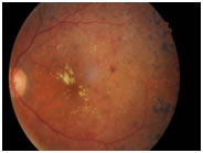
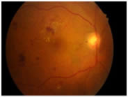
Dilated Eye Exam: During this part of the examination, the ophthalmologist or optometrist uses a special magnifying lens to examine you retina and optic nerve for signs of damage, such as leaking blood vessels, macular edema, fatty deposits, areas of ischemia, new blood vessel formation, and any change in shape to the blood vessels.
If the eye specialist suspects macular edema or leaking of vessels, additional testing may be performed. These tests include:
Fluorescein Angiography: In this test, a special dye is injected into your arm. As the dye passes through the blood vessels in your retina, pictures are taken. This test allows the ophthalmologist to find areas of leaking vessels and determine the best treatment.
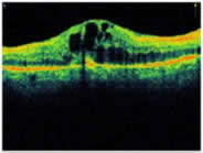
OCT: OCT, which stands for Optical Coherence Tomography, is a highly specific diagnostic test that allows the ophthalmologist to examine the cell layers of the retina and optic disc in detail. This diagnostic test is very useful in the diagnosis of macular edema.
What are the treatment options for diabetic retinopathy?
No treatment is typically needed during the first three stages of diabetic retinopathy (i.e. NPDR), unless macular oedema is present. The best way to prevent progression of diabetic retinopathy is to control levels of blood sugar, blood pressure, and cholesterol.
Macular Oedema:
Macular oedema can be treated with
Intravitreal injections: of Anti-VEGF (avastin) or Steroids (Triamcinolone / IVTA/ Ozurdex)
Focal Grid Laser : Your ophthalmologist will place up to several small laser burns in the areas of retinal leakage surrounding the macula. These burns slow the leakage of fluid and reduce the amount of fluid in the retina. Focal laser treatment stabilises vision. It can reduce the risk of vision loss by 50 percent. This treatment is usually completed in one session, however, further treatment may be needed.
Although both treatments have high success rates, they are not a cure for diabetic retinopathy.
Indications of endoscopic treatment:
Laser surgery (pan retinal photocoagulation / PRP / green laser)
Scatter Laser treatments shrink the abnormal blood vessels and prevent further bleeding. Your doctor places laser burns in the peripheral areas of the retina, causing the abnormal vessels to shrink. Due to the high number of laser burns, several treatment sessions are necessary.
Although you may notice some loss of your side vision, laser treatments can preserve the rest of your sight. Laser treatment may slightly reduce your color and night vision.
Vitrectomy
If there is severe bleeding in the retina, a vitrectomy may be performed. During a vitrectomy, blood is removed from the center of the eye. A vitrectomy is performed under local anesthesia. Your doctor makes are tiny incision in your eye and inserts a small instrument which is used to remove the vitreous gel that is clouded with blood. The vitreous gel is replaced with salt solution.
At Sharnam Eye Centre & Laser Services we perform minimal invasive vitreous surgery or Sutureless vitrectomy for complicated diabetic retinal problems.
Anti-VEGF
Avastin (Bevacizumab) is an anti-growth factor drug that is delivered through an intravitreal injection. This drug is used to reduce macular oedema associated with diabetes and also to reduce new vessel growth.
Usually, treatment helps slow or stop the progression of diabetic retinopathy. Treatment doesn’t cure the underlying disease. Careful diabetes management is important to reduce the risk of further eye damage.
PDR may cause more severe vision loss than NPDR because it can affect both central and peripheral vision. PDR affects vision in the following ways:
Vitreous hemorrhage: delicate new blood vessels bleed into the vitreous — the gel in the center of the eye — preventing light rays from reaching the retina. If the vitreous hemorrhage is small, you may see a few new, dark floaters. A very large hemorrhage might block out all vision, allowing you to perceive only light and dark. Vitreous hemorrhage alone does not cause permanent vision loss. When the blood clears, your vision may return to its former level unless the macula has been damaged.
Traction retinal detachment: scar tissue from neovascularization shrinks, causing the retina to wrinkle and pull from its normal position. Macular wrinkling can distort your vision. More severe vision loss can occur if the macula or large areas of the retina are detached.
Neovascular glaucoma: if a number of retinal vessels are closed, neovascularization can occur in the iris (the colored part of the eye). In this condition, the new blood vessels may block the normal flow of fluid out of the eye. Pressure builds up in the eye, a particularly severe condition that causes damage to the optic nerve.
Sharnam Eye Centre & Laser Services is a specialised centre having all facilities for the examination and medical or surgical treatment of diabetic eye disease.


