Why You Should Get an Eye Disease Screening at 40: Protect Your Vision as You Age

As we age, taking care of our health becomes more important, and that includes our eye health. The American Academy of Ophthalmology recommends that all adults get a baseline eye disease screening at age 40, even if you’ve never had vision problems before. This simple but essential step can help you maintain healthy vision for years to come.

Why Is Age 40 a Key Milestone for Eye Health?

By the time we reach 40, our eyes have undergone significant changes. Many common eye diseases, such as glaucoma, diabetic retinopathy, and cataracts, start to develop around this age, often without any noticeable symptoms. An eye screening at 40 provides a critical baseline for your eye health, allowing your ophthalmologist to detect early signs of disease and monitor changes over time.

What Happens During an Eye Disease Screening?

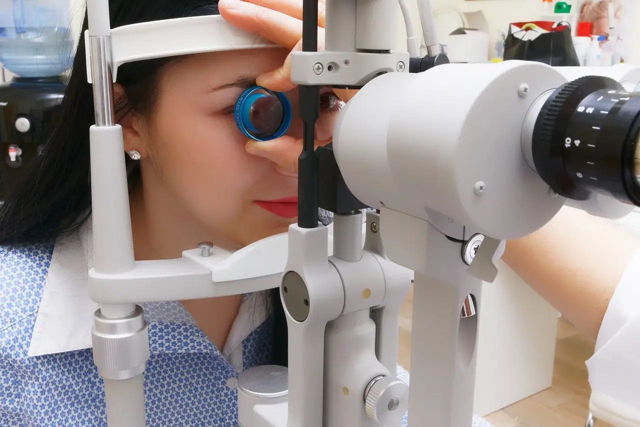
During your screening, an ophthalmologist will conduct a comprehensive examination of your eyes. This includes:

  • Visual Acuity Test: Checking how well you can see at various distances.
  • Eye Pressure Test: Measuring intraocular pressure to screen for glaucoma.
  • Pupil Dilation: Widening the pupils to examine the retina and optic nerve.
  • Visual Field Test: Assessing your peripheral vision, which can reveal issues like glaucoma.

These tests are painless and provide vital information about your eye health.

Common Eye Diseases Detected in People Over 40

  1. Glaucoma: Often called the “silent thief of sight,” glaucoma can lead to irreversible vision loss without any early symptoms. Early detection through an eye screening is key to managing this condition effectively.
  2. Diabetic Retinopathy: This condition is a leading cause of blindness in working-age adults and is closely linked to diabetes. Regular eye exams can catch diabetic retinopathy early, when treatment is most effective.
  3. Cataracts: While commonly associated with aging, cataracts can start to develop as early as 40. Regular eye exams help monitor the progression of cataracts, allowing for timely treatment to preserve vision.

Who Should Get an Eye Screening Before 40?

If you have certain risk factors, you shouldn’t wait until 40 for an eye screening. These risk factors include:

  • Diabetes
  • High blood pressure
  • Family history of eye disease
  • Existing eye conditions or injuries

People with these risk factors should see an ophthalmologist regularly, regardless of age.

Protect Your Vision: Schedule an Eye Screening Today

Think of an eye disease screening at 40 as you would other important health screenings, like mammograms or colonoscopies. It’s a proactive step to protect your vision and catch potential issues early, when treatment is most effective.

If you’re 40 or older and haven’t had a recent eye exam, now is the time to schedule one. Your eye health is too important to ignore, and a simple screening can make all the difference in preserving your vision for years to come.

Share the Importance of Eye Health

Taking care of your eyes is an essential part of overall health, and it’s something everyone should prioritize. Share this information with friends and family to encourage them to schedule their eye screenings as well. Together, we can help ensure that everyone enjoys clear, healthy vision throughout their lives.

For more information and to schedule your eye disease screening, contact your local ophthalmologist today.
Your vision matters—don’t wait to protect it.

Understanding Eye Twitching: Causes and Simple Remedies

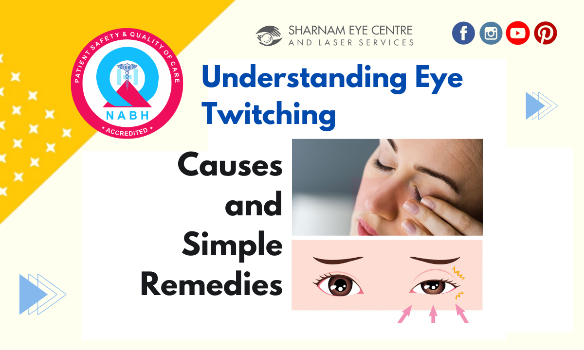
Eye twitching, also known as eyelid spasms or tics, is a common experience that many of us have faced at some point. It’s that sudden, involuntary movement of your upper or lower eyelid that can last for a few seconds, minutes, or even days. While it’s usually harmless, it can be quite annoying, especially if it persists.

Common Causes of Eye Twitching

  1. Sleep Deprivation: Not getting enough sleep is a common trigger for eye twitching. When you’re tired, your body may react by causing your eyelid muscles to spasm. The simple fix? Make sure you’re getting enough restful sleep each night.
  2. Stress: High levels of stress can also lead to eye twitching. If you’re feeling overwhelmed, it’s important to take steps to manage your stress. Relaxation techniques, such as deep breathing, meditation, or even a short walk, can help reduce your stress levels.
  3. Too Much Caffeine: Caffeine is a stimulant that can sometimes lead to eyelid spasms, especially if you’re consuming large amounts of coffee, tea, or soda. Cutting back on your caffeine intake might help alleviate the twitching.
  4. Dry Eyes: For some, dry or irritated eyes can cause twitching. If you experience dry eyes, using artificial tears or other treatments recommended by your eye doctor can provide relief.

When to Seek Help

In most cases, eye twitching is nothing to worry about and will go away on its own. However, if your eye twitching lasts for an extended period, becomes more frequent, or is accompanied by other symptoms, it’s a good idea to consult with a healthcare professional. They can rule out any underlying conditions that might be causing the issue.

By understanding the common causes of eye twitching and taking simple steps to address them, you can usually stop the twitch and get back to your day with ease.

A Memorable Homecoming: Dr. Puneet Gupta’s Talk at King George’s Medical University

As a proud alumnus of King George’s Medical University’s Department of Ophthalmology, it’s with great humility and joy that I share my recent experience of delivering a talk at my alma mater. The occasion was none other than the prestigious International Georgian Meet held on 23rd December 2024, a gathering that celebrates the excellence and heritage of our institution.

Receiving an invitation to present at the alma mater is always a moment of great honor, and for me, it was no different. I extend my heartfelt gratitude to Prof. and Head Dr. Apjit Kaur, along with all the esteemed faculty members of the Department, for this opportunity. The chance to engage with my former department, where my foundational journey in ophthalmology began, was both nostalgic and exhilarating.

My talk centered on the latest diagnostic modality for retinal disorders, OCT Angiography. This cutting-edge technology represents a significant leap in our ability to understand and treat various retinal conditions, and sharing insights on this topic with current students and faculty was an intellectually enriching experience.


Perhaps the most delightful aspect of this visit was the interaction with the present students of ophthalmology. Witnessing their enthusiasm, curiosity, and the sheer brilliance was not only uplifting but also a vivid reminder of my own days at the university. The vibrant academic environment of King George’s Medical University continues to foster a new generation of brilliant minds, and it was an honor to contribute, even if in a small way, to their educational journey.

As I reflect on the event, I am filled with a sense of deep connection and pride for my alma mater. The Department of Ophthalmology at King George’s Medical University is more than just an academic institution; it is a nurturing ground for medical pioneers and visionaries. To have been a part of this lineage and to continue being involved in its journey is a privilege I cherish immensely.

This day, marked by the exchange of knowledge and shared passion for ophthalmology, will remain etched in my memory. It was not just a return to a physical location but a reconnection with a fundamental part of my professional identity.

I look forward to many more such engagements in the future, where we can continue to build bridges between past, present, and future generations of medical professionals. Let us keep striving for excellence in our field and keep the Georgian spirit alive and thriving.

Dr. Puneet Gupta
Alumnus, Department of Ophthalmology
King George’s Medical University, Lucknow

FAQs about Retinal Detachment

Top 10 FAQs about Retinal detachment and its treatment – Understanding Retinal detachment

Top 10 FAQs about Retinal detachment and its treatment : Understanding Retinal detachment : A Comprehensive Guide to Treatment.

1. What is Retinal Detachment?

  • Retinal detachment occurs when the retina, the light-sensitive layer at the back of the eye, separates from its normal position. This can lead to vision loss and is considered a medical emergency.

2. What Causes Retinal Detachment?

  • Common causes include aging, eye injuries, certain eye surgeries, or disorders like diabetic retinopathy. It can also occur spontaneously due to changes in the vitreous gel inside the eye.

3. What are the Symptoms of Retinal Detachment?

  • Symptoms often include a sudden appearance of floaters (small specks or threads), flashes of light in one or both eyes, and a shadow or curtain effect over a portion of the visual field.

4. How is Retinal Detachment Diagnosed?

  • Diagnosis typically involves a comprehensive eye examination, including dilating the pupils to examine the retina. Special imaging tests, like ultrasound or optical coherence tomography (OCT), may also be used.

5. What are the Treatment Options for Retinal Detachment?

  • Treatment options include laser surgery, cryopexy (freezing), pneumatic retinopexy (injecting a gas bubble into the eye), scleral buckling, and vitrectomy. The choice depends on the type and severity of the detachment.

6. What is the Success Rate of Retinal Detachment Surgery?

  • The success rate for retinal detachment surgery is generally high, with about 85-90% of cases successfully treated with a single operation. Some cases might require additional procedures for complete resolution.

7. What are the Risks and Complications of Treatment?

  • Complications can include infection, bleeding, increased eye pressure, cataracts, or recurrence of the detachment. In rare cases, vision might not be fully restored.

8. How Long is the Recovery Process After Treatment?

  • Recovery time varies; it can take several weeks to months. Restrictions like avoiding certain activities or positioning the head in a specific way for a period are common, especially in surgeries involving gas bubbles.

9. Can Retinal Detachment be Prevented?

  • While not all cases can be prevented, regular eye exams, especially for those with risk factors, can lead to early detection and treatment. Protecting the eyes from injury and managing underlying conditions like diabetes also help.

10. What Lifestyle Changes are Recommended After Treatment?

  • After treatment, it’s important to avoid activities that could strain the eyes. This includes heavy lifting or vigorous exercise. Regular follow-up appointments are crucial. Patients should also be aware of the signs of re-detachment and seek immediate medical attention if they occur.

These responses offer a detailed overview of each aspect of retinal detachment and its treatment. It’s always advisable to consult with an eye care professional for personalized advice and treatment options.

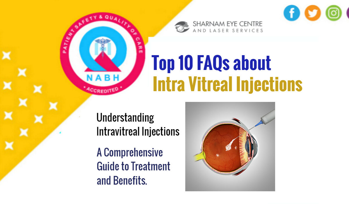
Understanding Intravitreal Injections

Top 10 FAQs about Intra Vitreal Injections – Understanding Intravitreal Injections

Top 10 FAQs about Intra Vitreal Injections: Understanding Intravitreal Injections: A Comprehensive Guide to Treatment and Benefits.

1. What is an Intravitreal Injection?

  • It’s a medical procedure where medication is injected into the vitreous, which is the jelly-like substance inside the eye, near the retina.

2. What Conditions are Treated with Intravitreal Injections?

  • These injections are primarily used to treat eye diseases such as age-related macular degeneration, diabetic retinopathy, and retinal vein occlusion.

3. How is the Intravitreal Injection Procedure Performed?

  • The eye is numbed with drops or an injection, the eye area is cleaned, and then the medication is injected into the vitreous. The procedure is usually quick, lasting only a few minutes.

4. Are Intravitreal Injections Painful?

  • Most patients experience little to no pain due to the numbing of the eye, but some might feel pressure or a slight sting.

5. What are the Risks and Side Effects of Intravitreal Injections?

  • Possible risks include infection, bleeding, increased eye pressure, cataracts, and in rare cases, retinal detachment.

6. How Effective are Intravitreal Injections?

  • Effectiveness varies, but these injections can significantly improve or stabilize vision for many patients, especially when started early in the disease process.

7. How Often Will I Need Intravitreal Injections?

  • The frequency depends on the condition and response to treatment. Initially, injections might be monthly, with adjustments based on how the eye responds.

8. What Should I Expect After the Injection?

  • Temporary side effects may include redness, floaters, or eye discomfort. Normal activities can usually be resumed shortly after, but your doctor may advise against strenuous activities for a few days.

9. How Should I Prepare for an Intravitreal Injection?

  • No specific preparation is usually required, but follow any instructions given by your doctor, such as avoiding certain medications or using antibiotic drops.

10. Are There Alternative Treatments to Intravitreal Injections?

  • Alternatives or complementary treatments may include laser therapy, oral medications, or lifestyle changes, depending on the condition being treated.

Always consult with a healthcare provider for information and advice specific to your situation.

FAQ for Age-Related Macular Degeneration

Top 10 FAQ for Age-Related Macular Degeneration – Understanding ARMD or AMD

Top 10 FAQ for Age-Related Macular Degeneration – Understanding ARMD or AMD: Symptoms, Risks, and Treatments – A Comprehensive Guide

1. What is age-related macular degeneration?

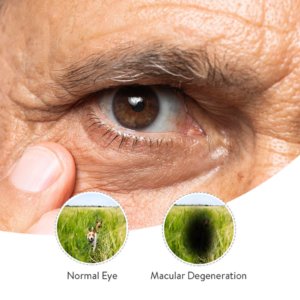
AMD is a common eye condition and a leading cause of vision loss among people age 50 and older. It causes damage to the macula, a small spot near the center of the retina and the part of the eye needed for sharp, central vision.

2. What are the types of AMD?

There are two main types: Dry AMD, which is more common and less severe, and Wet AMD, which is more serious and can lead to more rapid vision loss.

3. What are the symptoms of AMD?

Early AMD often doesn’t have symptoms. Later stages may include blurred or reduced central vision, straight lines appearing wavy, and difficulty seeing in low light.

4. Who is at risk for AMD?

Risk factors include age (over 50), smoking, genetics, race (Caucasians are more likely to develop AMD than other races), and family history.

5. How is AMD diagnosed?

AMD is diagnosed through a comprehensive eye exam that includes visual acuity testing, a dilated eye exam, and sometimes optical coherence tomography (OCT) or fluorescein angiography.

6. Can AMD be prevented?

While AMD cannot be prevented, risk can be reduced by not smoking, eating a healthy diet rich in green, leafy vegetables and fish, maintaining normal blood pressure and cholesterol levels, and having regular eye exams.

7. How is AMD treated?

Treatment depends on the type of AMD. For dry AMD, there’s no current treatment but lifestyle changes can slow progression. For wet AMD, treatments include anti-VEGF injections, laser therapy, and photodynamic therapy.

8. What are anti-VEGF injections?

Anti-VEGF therapy involves injections into the eye to block a protein that stimulates the growth of abnormal blood vessels in the retina. It’s a common treatment for wet AMD.

9. Is there a cure for AMD?

Currently, there is no cure for AMD, but treatments can slow the progression or improve vision.

10. What is the outlook for people with AMD?

The outlook varies depending on the type and stage of AMD. Many people maintain active lifestyles despite AMD. Regular eye care and treatments can help manage the condition and preserve vision.

It’s important for individuals to have regular eye examinations, especially those over 50, to detect AMD early and manage its progression effectively.

Disclaimer: This content is for informational purposes only and should not replace professional medical advice, diagnosis, or treatment.

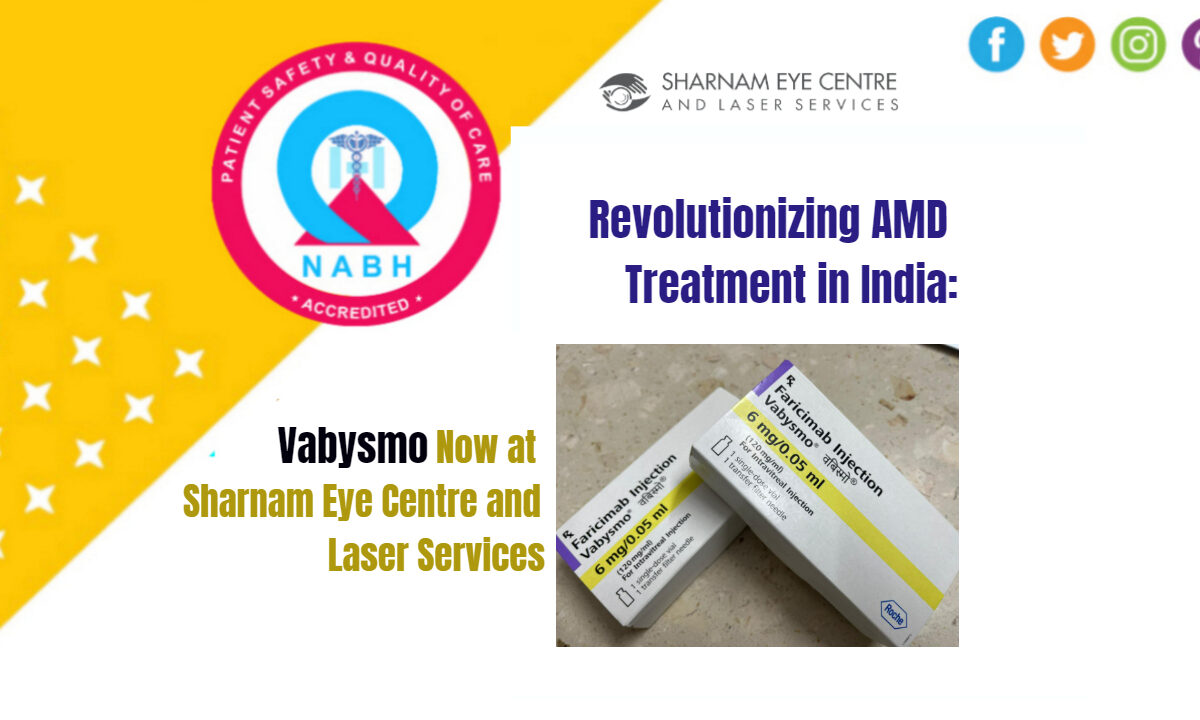
Vabysmo injection

Revolutionizing AMD Treatment in India: Vabysmo Now at Sharnam Eye Centre and Laser Services

Introduction:

Welcome to a new era in eye care! At Sharnam Eye Centre and Laser Services in Ghaziabad, we’re excited to introduce Vabysmo, a groundbreaking treatment for Age-Related Macular Degeneration (AMD). This latest advancement is a beacon of hope for individuals struggling with this challenging condition. Our blog today delves into how Vabysmo injection is transforming the AMD treatment landscape in India.

Understanding AMD and Its Impact:

Understanding AMDAMD, primarily affecting the elderly, is a major cause of vision loss. It damages the macula, a crucial part of the retina, leading to blurred or no vision in the center of the visual field. Traditional treatments have centered around targeting Vascular Endothelial Growth Factor (VEGF), a key player in the development of wet AMD. However, with evolving medical science, new treatments like Vabysmo are emerging, offering a more comprehensive approach.

Vabysmo: A Dual-Action Treatment:

Vabysmo, known scientifically as faricimab, is an FDA-approved drug that marks a significant advancement in AMD treatment. Unlike earlier medications focusing solely on VEGF, Vabysmo targets both VEGF and Angiopoietin-2. This dual-action approach addresses two fundamental causes of AMD, offering a more potent defense against this eye condition.


Vabysmo InjectionBenefits of Vabysmo at Sharnam Eye Centre:
  1. Longer Lasting: One of the standout features of Vabysmo is its extended effectiveness. Patients may only need treatment once every three to four months, reducing the frequency of hospital visits.
  2. Comprehensive Treatment: By targeting two key causes of AMD, Vabysmo injection offers a more thorough treatment approach, potentially improving outcomes for our patients.
  3. Expert Care: At Sharnam Eye Centre, our team of specialists is trained in the latest ophthalmologic procedures, ensuring you receive top-notch care with Vabysmo.

Why Choose Sharnam Eye Centre?

Choosing the right care center is crucial. At Sharnam Eye Centre, we blend advanced technology with compassionate care to provide the best treatment experience. Our state-of-the-art facilities and patient-centered approach make us a leading choice for eye care .

Conclusion:

The introduction of Vabysmo at Sharnam Eye Centre represents a significant step forward in AMD treatment . With its dual-action formula and longer-lasting effects, it offers a new hope for those affected by this condition. We are committed to providing our patients with the latest and most effective treatments available.

If you or a loved one is dealing with AMD, don’t wait. Contact Sharnam Eye Centre today to learn more about how Vabysmo can help you reclaim your vision and quality of life.

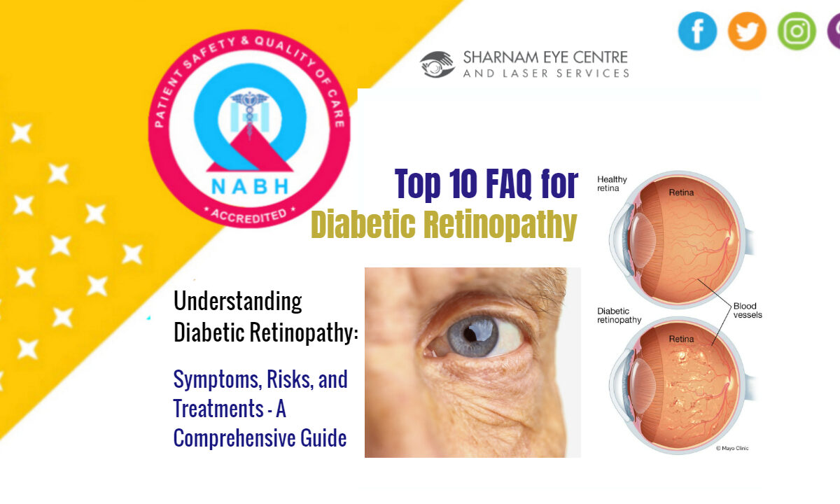
FAQ on Diabetic Retinopathy

Top 10 FAQ for Diabetic Retinopathy – Understanding Diabetic Retinopathy

Top 10 FAQ for Diabetic Retinopathy – Understanding Diabetic Retinopathy: Symptoms, Risks, and Treatments – A Comprehensive Guide

1. What is diabetic retinopathy?

Diabetic retinopathy is a diabetes complication that affects the eyes. It’s caused by damage to the blood vessels of the light-sensitive tissue at the back of the eye (retina).

2. What are the symptoms of diabetic retinopathy?

In the early stages, diabetic retinopathy may cause no symptoms or only mild vision problems. Eventually, it can cause blindness. Symptoms may include spots or dark strings floating in your vision (floaters), blurred vision, fluctuating vision, dark or empty areas in your vision, and vision loss.

3. Who is at risk for diabetic retinopathy?

Anyone with type 1 or type 2 diabetes can develop this condition, especially if they have had diabetes for a long time, have poorly controlled blood sugar, high blood pressure, high cholesterol, or are pregnant.

4. How is diabetic retinopathy diagnosed?

It’s diagnosed during an eye examination that includes visual acuity testing, dilating the eyes to examine the retina and optic nerve, and possibly fluorescein angiography or optical coherence tomography (OCT).

5. Can diabetic retinopathy be prevented?

Managing your diabetes is the best way to lower your risk. This includes controlling your blood sugar level, blood pressure, and cholesterol, along with regular diabetes and eye check-ups.

6. What are the treatments for diabetic retinopathy?

Treatment, which can’t cure diabetic retinopathy, often slows or stops the progression of the condition. Treatments may include laser treatment, injection of corticosteroids or anti-VEGF medication into the eye, and vitrectomy surgery.

7. Is laser treatment for diabetic retinopathy painful?

Most patients experience little to no discomfort during laser treatment. You may feel a sharp prick or a sensation of pressure in the eye. The eye is usually numbed before treatment.

8. What are the side effects of treatments for diabetic retinopathy?

Side effects can include bleeding in the eye, increased pressure in the eye, pain or discomfort in the eye, and temporary or permanent loss of vision.

9. How often should someone with diabetes get an eye exam?

If you have diabetes, it’s recommended to have a comprehensive dilated eye exam at least once a year. More frequent medical eye exams may be necessary if you have diabetic retinopathy or if your eye doctor advises it.

10. Can vision loss from diabetic retinopathy be reversed?

Unfortunately, loss of vision from diabetic retinopathy is often irreversible. However, early detection and treatment can significantly reduce the risk of blindness and can sometimes restore vision that has been lost.

Disclaimer: This content is for informational purposes only and should not replace professional medical advice, diagnosis, or treatment.

Understanding Intra Vitreal Injections at Sharnam Eye Centre and Laser Services

Introduction:
Intra Vitreal injections are a cornerstone in the treatment of various retinal diseases. At Sharnam Eye Centre and Laser Services, under the expert guidance of Dr. Puneet Gupta, a vitreo-retinal specialist since 1993, these injections are administered with precision and care.

What are Intra Vitreal Injections?

Intra Vitreal injections involve the delivery of medication directly into the vitreous, the jelly-like substance inside the eye. This method is primarily indicated for conditions like age-related macular degeneration, diabetic retinopathy, retinal vein occlusion, and endophthalmitis.

Procedure at Sharnam Eye Centre:

Dr. Gupta and his team ensure the process is as comfortable as possible. The eye is numbed with topical anesthesia, making the procedure virtually painless. Using state-of-the-art equipment, the medication is precisely injected, minimizing any discomfort.

Post injection care:

After the injection, patients might experience slight discomfort or redness, which is normal. The team at Sharnam Eye Centre provides detailed after-care instructions, emphasizing the importance of not rubbing the eye and watching for signs of complications like pain or decreasing vision.

Why Choose Sharnam Eye Centre?

  1. Expertise: Dr. Gupta’s extensive experience since 1993 in vitreo-retinal diseases ensures expert handling of intricate procedures.
  2. Advanced Technology: The centre is equipped with the latest technology for precise and effective treatment.
  3. Patient-Centric Approach: Personalized care and attention to each patient’s specific needs are paramount.
  4. Comprehensive Care: Beyond injections, the centre offers a full range of services for retinal and vitreous diseases, ensuring continuity of care.
  5. Educational Focus: Patients receive detailed information about their condition and the treatment, empowering them with knowledge about their eye health.

Conclusion:

Intra Vitreal injections can be a crucial treatment for retinal diseases. At Sharnam Eye Centre, patients receive top-notch care under Dr. Puneet Gupta’s expertise. The combination of advanced technology, a patient-centered approach, and comprehensive care makes Sharnam Eye Centre a premier choice for those seeking intra vitreal treatments.

Note: Always consult with a healthcare professional for personal medical advice.

1 2 3 6
Appointment
close slider

For Appointment-

Online Appointment: Available between 8 AM to 8 PM (Except Saturday)

Book An Online Appointment - Click Here

Call for Appointment -

9818851925,

9810979457,

8586030104

-----------------------------------------